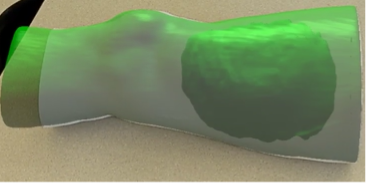
For neoadjuvant radiation therapy treatment for Sarcoma, radiation oncologists prepare radiation dose distribution maps based off of CT scans. After administration of radiation therapy, often a tumor resection is performed for sarcoma patients who are in a later stage of cancer. To mitigate tissue necrosis following tumor resection surgery, surgical oncologists would benefit from viewing the radiation dose distribution on the anatomy of the patient. Through prescribing feature points and the camera input from an augmented reality headset, the virtual model of the patient’s radiation dose distribution can be overlayed onto their physical anatomy in the operating room via means of an augmented reality application. During the augmented reality application runtime, the alignment of the virtual hologram to the physical anatomy is automatic, in real-time, and stable across multiple viewing angles. In addition, users can manipulate aspects of the virtual model with thresholds of radiation, transparency, and toggleable regions.